
By Brittney M. McInnis, Tyler W. Hodges,* Lisa K. Kemp, Jonathan D. Hurt, and Steve McDaniel, Reactive Surfaces and Aayushma Kunwar,** William Carey University
The antimicrobial properties of two disparate bio-based coating additives were evaluated in a polyvinyl alcohol (PVA) food packaging coating for antimicrobial activity. Chitosan, a shrimp and crustacean shell derived polysaccharide, and an antimicrobial peptide were evaluated in a dissolvable food package coating for reductions in microbial growth after contacting agar patties serving as food simulants. Where the antimicrobial components of such packaging coatings are chosen to be generally recognized as safe by worldwide regulatory agencies, migration from the packaging into headspaces and food-contact surfaces can provide enhanced efficacy against foodborne pathogens, including viruses. The techniques and coatings presented in this article suggest that dramatic improvements in food safety can be achieved using coatings containing non-toxic bio-based biocides.
Introduction
Food packaging is an asset that can preserve foods going to market and extend shelf-life, but it can also become a liability when the packaged food itself is contaminated or becomes so during the packaging process. The economic damage to the food industry of reduced shelf-life for packaged foods is great, and contamination of packaged food is a major public health concern. There are several contamination points during food preparation and packaging and numerous examples of outbreaks of foodborne illness leading to the recall of goods.1-3 Major incidences have occurred around the world over the decades (e.g., E. coli O157:H7 contamination from the United States Jack in the Box restaurant,4 E. coli O104:H4 spread from German sprouts,5 listeriosis throughout Europe in frozen corn,6 E. coli O157:H7 on Canadian pork,7 listeriosis from processed meat in South Africa8) and continue today. Table 1 highlights the diversity of products and pathogens associated with some of the most recent outbreaks in the United States.9 Even as recently as July 2018, there was a recall of packaged vegetable trays due to a multistate outbreak of parasitic cyclosporiasis in the United States.10

Such outbreaks have led not only to changes in guidelines and adoption of new regulations,11-13 but also spurred interest in new technologies such as “smart” packaging.14,15 Packaging coatings that can reduce such contamination while the food is traveling to its point of sale are long-sought goals of the food industry. Research for improved food packaging has generally focused on two areas: edible films that can be applied directly to food and are safe to eat,16-19 and chemical or physical modifications to plastic packaging materials that are permanent and specifically do not leach onto or into the packaged food product.20-22 In this work, we combined the best attributes of both research areas into one product by retaining the physical barrier properties afforded by plastic packaging film and gaining the effectiveness of dissolvable (but safe to eat) bio-based antimicrobial additives. Bio-based additives have the advantage of often having low toxicity to humans, and many have notifications with the Food and Drug Administration that they have been determined to be Generally Recognized as Safe (GRAS). Though not a prerequisite for development of an antimicrobial food contact coating in the present study, the selection of materials, whether polymeric coating components or bio-based additives, with GRAS notices previously filed was a consideration. Both polyvinyl alcohol and chitosan fall into this category and were chosen for this study.23-26 Other considerations were the likely efficacy of the additive against microorganisms commonly associated with foodborne illness (i.e., bacteria like Escherichia coli), known general lack of toxicity, and biodegradability. Finally, as the numbers and types of foodborne pathogens are varied, consideration was given to bio-based biocides that were potentially capable of controlling not only bacteria, but also spores of Gram-positive species, fungi, algae, and viruses (ergo, the inclusion of antimicrobial peptides).
Two quantitative assays were developed to analyze effectiveness of the coatings using modern microbiological methods and statistical software. The first used clear-coated plastic disks through which bacterial colonies could be enumerated on the agar surface beneath. The second used agar “patties” serving as a food simulant in a vacuum-sealed food packaging system. The contaminated agar can be contacted on one or both sides with an antimicrobial coating and the microbial growth inhibition evaluated. Statistical evaluation was undertaken to detect antagonistic, additive, and/or synergistic relationships between chitosan and an antimicrobial peptide, AMP7, in the food contact coating.27-29 The assays were successful in detecting antimicrobial activity against E. coli for both the bio-based additives analyzed, and the statistical methods used detected an antagonistic effect at most of the concentrations evaluated and only slightly antagonistic or additive effects at higher concentrations. These methods proved useful in screening the candidate bio-based additives presented here and are currently being used to evaluate other promising bio-based additives for incorporation into food packaging.
Materials and Methods
Reagents and Bacterial Strains
Polyvinyl alcohol (PVA) was obtained from Sigma-Aldrich (Cat# 348406, reported Mw 13,000-23,000, 98% hydrolyzed). Chitosan was obtained from bulksupplements.com. E. coli K12 was obtained from Presque Isle Cultures (Erie, PA), and the peptide AMP7 was obtained from Reactive Surfaces, Ltd. (Austin, TX). Isopropanol (91%) was purchased locally. All growth media used Difco Tryptic Soy Agar (TSA) or tryptic soy broth from Becton, Dickinson, and Co. (Sparks, MD). MacConkey, Eosin Methylene Blue (EMB), and Luria-Bertani (LB) agar were obtained from Carolina Biological Supply Co. (Burlington, NC). 4-Methylumbelliferyl-β-D-glucuronide dehydrate (MUG) was obtained from Fisher Scientific (Waltham, MA).
Preparation of Coated Films
Disks of 0.5-in. diameter were cut using a 40W CO2 laser (Glowforge®, Seattle, WA) from a 3-mil thick sheet of clear Dura-Lar polyester (Grafix, Maple Heights, OH) substrate. For the vacuum-sealed food simulant experiment, 8.5 cm diameter disks were hand-cut from Dura-Lar sheets, or from commercial substrates, including Opalen™ (Bemis, Parc de L’Alliance Braine L’Alleud, Belgium), a clear, PET film and Trayforma™ (Stora Enso, Stockholm, Sweden), a PET-coated paperboard. A 5% (w/w) PVA solution, 1% (w/w) chitosan in 2% (v/v) acetic acid solution, and 10% (w/w) AMP7 in 5% (w/w) PVA solution were prepared and mixed before application to create the final concentrations indicated in the following experiments. In each case, the bio-based additive levels reported are based on the percentage by weight in the final, dry coating. All Dura-Lar films were coated by applying a specific volume of the liquid coating directly to the film, so that the final film thickness was approximately 1 mil. The coated films were left to dry at room temperature overnight before being used in any of the antimicrobial tests.
Antimicrobial Coated Disk Treatment of E. coli Contaminated Agar Plates
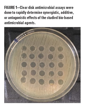
Traditional zone of inhibition testing uses paper disks infused with the target active. The paper disks are incubated with microbes, and the zone of clearing seen around the disk (i.e., lack of microbial colony growth due to leaching of the active from the disk) is measured to indicate the effectiveness of the active. In our experiments, using the transparent disks with a clear, dissolvable coating allowed inspection of cell growth directly beneath the disk as well as any observable zone of inhibition (Figure 1). A cotton swab was dipped into a suspension of E. coli K12 (~5×10⁶ CFUs/mL) and was spread over 15 cm diameter TSA plates (done in triplicate). After the plates had dried for approximately 15 min, the disks were placed, coated side down, on top of the E. coli layer. The plates were incubated at 30°C to 36°C overnight. Each disk was photographed using a dissecting microscope for magnification, and colonies were counted using ImageJ software from the National Institutes of Health (Bethesda, MD). The dose response of individual additives was evaluated using log-logistic regression model with the R package drc.30 For experiments involving the combination of bio-based additives in coatings, the interaction between the two bioadditives (i.e., synergistic, additive, antagonistic) was evaluated using the zero interaction potency (ZIP) model with the R package synergyfinder.31
Preparation of Vacuum-Sealed Food Simulants
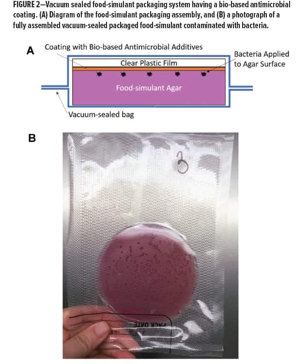
To mimic packaged food, agar patties were cast into petri dishes and then gently removed from the dishes once firm to serve as food-patty simulants. These patties were vacuum sealed with plastic film inserts containing mixtures of PVA, chitosan, and AMP7 as studied in the small disk assays (Figure 2). Several selective and differential agar media were evaluated for visualization of E. coli colonies. These included MacConkey agar, LB agar with MUG, and EMB agar. It was determined that MacConkey agar consistently produced clearly visible and easily observable E. coli colonies, and thus was primarily used. Each agar patty was placed in a vacuum bag, and an aliquot of diluted E. coli was spread over one surface so that approximately 200 CFUs were added (each test done in triplicate). Coated films sized to match the agar patties were placed, coating side facing the bacteria, on the agar patties, and uncoated films were used as controls. The vacuum seal bags were sealed using the “Low” vacuum setting and the default seal setting on a Harvest Keepers Commercial vacuum sealer. The vacuumed samples were incubated for 24 h at 30°C, and colonies were counted using ImageJ software. For experiments measuring the effect of AMP7 and chitosan combinations, the interaction between the two bioadditives (i.e., synergistic, additive, antagonistic) was evaluated using the R package synergyfinder, with the Bliss model being used to calculate predicted response because the number of combinations was low.
Experiments and Results
Clear Disk Antimicrobial Assay
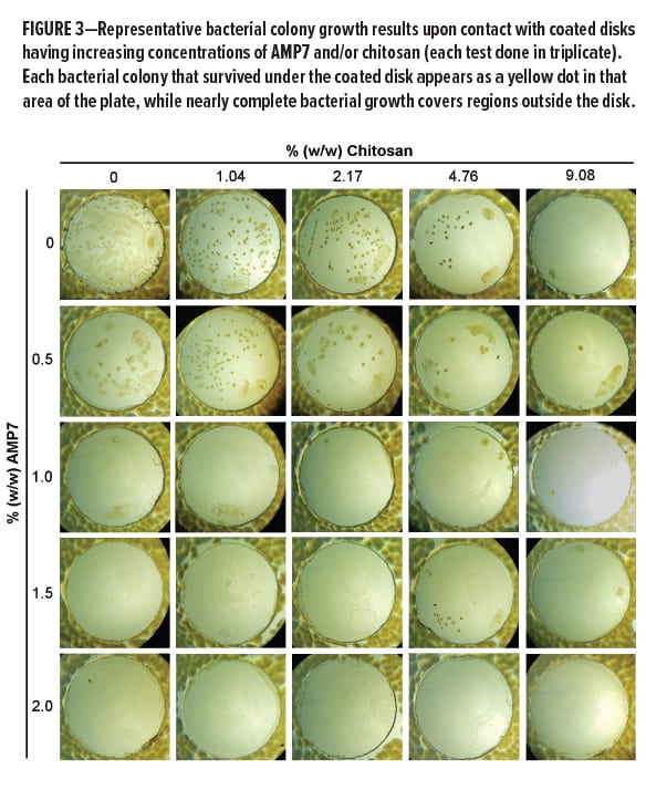
The efficacy of the bio-based antimicrobials in food-safe coatings was assessed by placing the coated 0.5-in. diameter Dura-Lar disks, coating-side down, onto prepared lawns of E. coli (Figure 3). The disks were coated with PVA-based coatings dosed with AMP7 at concentrations from 5,000 [0.5% (w/w)] to 20,000 ppm [2% (w/w)], and with chitosan at concentrations from 10,400 [1.04% (w/w)] to 90,800 ppm [9.08% (w/w)], or combinations of these two additives.
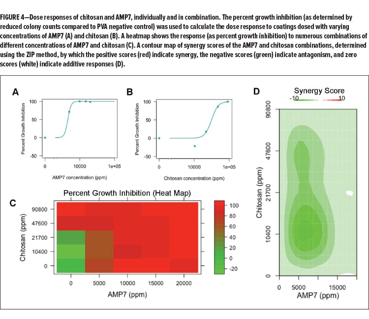 The dose response, as determined by percent reduction in colony numbers compared to the PVA negative control, was determined for AMP7 and chitosan. The effective dose to kill 50% of the bacterial population (ED50) of AMP7 was around 4,500 ppm and 30,000 ppm for chitosan (Figures 4A and 4B). The responses of the bacteria to various combinations are displayed in the heatmap (Figure 4C), which shows the relative response as a color from red (highest percent growth inhibition) to green (lowest percent growth inhibition). To test whether antagonism or synergy exists between these two compounds, the responses from AMP7 and chitosan combinations were used to determine the synergy score using the ZIP method, which returns a score based on the deviation of the actual response from the expected response.32 These scores are visualized for each combination in a contour plot (Figure 4D), which suggests that the two antimicrobials generally interact in an antagonistic manner, with the highest regions of antagonism existing for concentrations of AMP7 between 5,000 ppm [0.5% (w/w)] and 10,000 ppm [1.0% (w/w)] and for concentrations of chitosan ranging from 10,400 ppm [1.04% (w/w)] to 47,600 ppm [4.76% (w/w)]. However, beyond this region of antagonism, all other combinations of AMP7 and chitosan appear to interact with reduced levels of antagonistic behavior. This suggests that the AMP7 peptide and chitosan are interfering with each other’s function, but that the negative interaction can be overcome as the concentrations of both additives increase. The antagonistic response could be happening because both additives target the cellular membrane of microbes to produce a biocidal effect.
Moist-Food Simulant Packaging Study
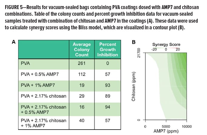
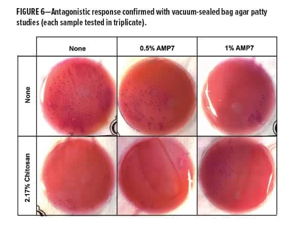
To test the scalability of the results from the clear disk assay, we simulated the common vacuum-sealed storage of moist foods (meats, fruits, vegetables, etc.) using a nutrient agar patty contacting a bio-based antimicrobial coated plastic disk while stored inside a vacuum sealed bag. Single additives were tested as well as combinations at corresponding concentrations. These results are summarized in the table in Figure 5A. Addition of 1% (w/w) AMP7 to 2.17% (w/w) chitosan-PVA coatings markedly worsened the efficacy of chitosan at this concentration, suggesting stronger antagonistic behavior in this concentration regime, which is consistent with the original model from the small disk study. Interaction between the two additives was evaluated by calculating synergy scores, using the Bliss model to calculate predicted responses. The contour plot of these scores is shown in Figure 5B. Although the combination of 0.5% (w/w) AMP7 with 2.17% (w/w) chitosan-PVA coating exhibited improved antimicrobial activity compared with the 0.5% AMP7 or 2.17% chitosan PVA coatings alone, the percent growth inhibition was less than predicted for an additive combination response, resulting in a negative synergy score (Figure 5B). Representative plates for this combination, as well as for the single additives and control, are shown in Figure 6.
 Commercial Packaging Study at Elevated Concentrations and Reduced Coating Thickness
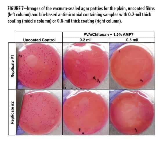
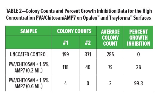
The vacuum-sealed patty studies had good agreement with the results seen in the disk assay, which confirmed that the disk assay is a good method for screening the effectiveness of these bio-based additives alone and in combination. To complete our study with this combination of materials, we wanted to overcome the antagonistic effects of these two components by increasing the final concentration in the PVA coating to 16% (w/w) chitosan and 1.5% (w/w) AMP7, and evaluating if cost-effective materials could be made by reducing the overall coating thickness on the test films. Because the bioactive coatings used in these studies are soluble, dosage of the active ingredients can be varied both by their concentrations in the coating mixture and by the amount of the coating added to the films (higher volumes applied to the films resulted in thicker coatings and a higher dose of actives). This test was conducted using disks of commercial packaging products. Disks of both types of commercial packaging were tested with either a 0.2-mil thick coating or a 0.6-mil thick coating (compared to the 1-mil thick coatings used in the earlier studies). As seen in Figure 7, it was confirmed that the bio-based additives could be used with commercial packaging materials to get efficient kill of E. coli contamination by controlling coating thickness to achieve enough of the chitosan and AMP7 to overcome their antagonistic effects (see Table 2).
 Conclusions
Individually, and in combination, AMP7 and chitosan in a simple PVA coating demonstrated effective antimicrobial activity in reducing bacterial growth in a food simulant contacting the food packaging coating. It was determined that combinations of AMP7 and chitosan had an antagonistic interaction, rather than additive or synergistic activity. This was not unexpected, as these additives, though biochemically dissimilar with one being a polysaccharide and the other an amino acid oligomer, act upon the same cellular target (the external cell membrane of bacteria and other microorganisms). When combined, they may compete for physical interaction with the cellular membrane to disrupt the membrane and produce a biocidal effect; so it is possible that one interfered with the other’s effectiveness. The antagonistic effects were shown to be overcome at high concentrations of both additives, and they are both still good candidates for the development of antimicrobial food packaging systems because they have demonstrated antimicrobial activity and are known to possess low toxicity; for example, chitosan has previously been used in antimicrobial edible coatings33,34 and the antimicrobial peptide used has previously been demonstrated to exhibit no discernable toxicity in rodent oral administration evaluations.35
The analytical methods used here offer a powerful tool for screening potential bio-based actives for additive, antagonistic, or synergistic activity. Other bio-based antimicrobials can be selected to act on different cellular targets, and combinations that target different cellular components would likely produce additive or synergistic antimicrobial effects. Selection of antimicrobials that interact synergistically in combination is ideal, because this increases the antimicrobial activity of both additives while decreasing the concentration needed, thereby reducing overall production costs in a commercial application. Enzymatic additives may be selected to catalyze destructive reactions on lipids, proteins, sugars, and cellular wall components that sustain microbial life. Enzymes can be selected to be specific to the biochemistry of target microorganisms, such as selecting an enzyme that preferentially degrades bacterial cell walls vs the cell walls of fungi. Alternatively, some enzymatic additives could be selected to exert nonspecific antimicrobial effects, such as certain oxidases that produce reactive oxygen species that attack most microbial biomolecules, including DNA. Other non-enzymatic peptide bio-additives, such as nisin and AMP7, have varying modes of action, typically through disrupting microbial membranes and cell walls, but due to their small molecular sizes, may be more suitable for applications where diffusion from a food preservative coating may aid in getting better protective coverage of the food item during storage. Future studies using different combinations of these types of bio-additives may produce coatings to safely enhance the shelf-life of food, and in some cases, be tailored to protection of specific food items from microbes, particularly pathogens, that preferentially contaminate those products.
References
- Carney, E., O’Brien, S.B., Sheridan, J.J., McDowell, D.A., Blair, I.S., and Duffy, G., “Prevalence and Level of Escherichia coli O157 on Beef Trimmings, Carcasses and Boned Head Meat at a Beef Slaughter Plant,” Food Microbiol., 23 (1), 52-59 (2006).
- Møretrø, T. and Langsrud, S., “Residential Bacteria on Surfaces in the Food Industry and Their Implications for Food Safety and Quality,” Comprehensive Reviews in Food Science and Food Safety, 16 (5), 1022-1041 (2017).
- Jay, M.T., Cooley, M., Carychao, D., Wiscomb, G.W., Sweitzer, R.A., Crawford-Miksza, L., Farrar, J.A., Lau, D.K., O’Connell, J., Millington, A., and Asmundson, R.V., “Escherichia coli O157: H7 in Feral Swine Near Spinach Fields and Cattle, Central California Coast,” Emerging Infectious Diseases, 13 (12), 1908 (2007).
- Pennington, H., Escherichia coli O157. The Lancet, 376 (9750), 1428-1435 (2010).
- Frank, C., Werber, D., Cramer, J.P., Askar, M., Faber, M., an der Heiden, M., Bernard, H., Fruth, A., Prager, R., Spode, A., and Wadl, M., “Epidemic Profile of Shiga-toxin–producing Escherichia coli O104: H4 Outbreak in Germany,” New Engl. J. Med., 365 (19), 1771-1780 (2011).
- Listeria monocytogenes: Update on Foodborne Outbreak. https://www.efsa.europa.eu/en/press/news/180703.
- Honish, L., Punja, N., Nunn, S., Nelson, D., Hislop, N., Gosselin, G., Stashko, N., and Dittrich, D., “Escherichia coli O157: H7 Infections Associated with Contaminated Pork Products—Alberta, Canada, July–October 2014,” Canada Communicable Disease Report= Releve Des Maladies Transmissibles Au Canada, 43 (1), 21-24 (2017).
- World Health Organization. Listeriosis–South Africa
- Centers for Disease Control and Prevention. List of Selected Multistate Foodborne Outbreak Investigations. https://www.cdc.gov/foodsafety/outbreaks/multistate-outbreaks/outbreaks-list.html.
- Del Monte Fresh Produce N.A., Inc. Voluntarily Recalls Limited Quantity of Vegetable Trays in a Multistate Outbreak of Cyclospora Illnesses in Select Retailers in Illinois, Indiana, Iowa, Michigan, Minnesota,
and Wisconsin, Because of Possible Health Risk. https://www.fda.gov/Safety/Recalls/ucm610992.htm. - Bennett, S.D., Sodha, S.V., Ayers, T.L., Lynch, M.F., Gould, L.H. and Tauxe, R.V., “Produce-associated Foodborne Disease Outbreaks, USA, 1998–2013,” Epidemiol. Infect., 1-10 (2018).
- Littlefield, R.S., “Jack in the Box: Lessons Learned by Accepting Responsibility,” Lessons Learned About Protecting America’s Food Supply, 35 (2005).
- Swanger, N. and Rutherford, D.G., “Foodborne Illness: The Risk Environment for Chain Restaurants in the United States,” Int. J. Hosp. Manage., 23 (1), 71-85 (2004).
- Malhotra, B., Keshwani, A. and Kharkwal, H., “Antimicrobial Food Packaging: Potential and Pitfalls,” Front. Microbiol., 6, 611 (2015).
- Vanderroost, M., Ragaert, P., Devlieghere, F. and De Meulenaer, B., “Intelligent Food Packaging: The Next Generation,” Trends in Food Sci. & Technol., 39 (1), 47-62 (2014).
- Embuscado, M. and Huber, K.C. (Eds.), Edible Films and Coatings for Food Applications. New York: Springer, 2009.
- Sapper, M. and Chiralt, A., “Starch-Based Coatings for Preservation of Fruits and Vegetables,” Coatings, 8 (152); DOI:10.3390/coatings8050152 (2018).
- Yu, D., Regenstein, J.M., and Xia, W., “Bio-based Edible Coatings for the Preservation of Fishery Products: A Review,” Crit. Rev. Food Sci. Nutrition, DOI: 10.1080/10408398.2018.1457623 (2018).
- Zimoch-Korzycka, A. and Andrzej Jarmoluk, A., “Polysaccharide-Based Edible Coatings Containing Cellulase for Improved Preservation of Meat Quality during Storage,” Molecules, 22 (390); DOI:10.3390/
molecules22030390 (2017). - Appendinia, P. and Hotchkiss, J.H., “Review of Antimicrobial Food Packaging,” Innov. Food Sci. Emerg. Technol., 3 (2), 113-126 (2002).
- Ahmed, I., Lin, H., Zou, L., Brody, A.L., Li, Z., Qazi, I.M., Pavase, T.R., and Liangtao Lv, L., “A Comprehensive Review on the Application of Active Packaging Technologies to Muscle Foods,” Food Control, 82, 163-178 (2017).
- Wang, H., Wei, D., Ziaee, Z., Xiao, H., Zheng, A., and Zhao, Y. “Preparation and Properties of Nonleaching Antimicrobial Linear Low-Density Polyethylene Films,” Ind. & Eng. Chem. Res., 54 (6), 1824-1831; DOI: 10.1021/ie504393t (2015).
- Agency Response Letter GRAS Notice No. GRN 000141 from George H. Pauli, Acting Director, Office of Food Additive Safety, Center for Food Safety and Applied Nutrition, FDA, to David R. Schoneker (on behalf of Colorcon) (April 28, 2004),
- GRAS Notice No. GRN 000397 from Dennis M. Keefe, Director, Office of Food Additive Safety, Center for Food Safety and Applied Nutrition, FDA, to Véronique Maquet (on behalf of KitoZyme S.A.) (Dec. 19, 2011)
- GRAS Notice No. GRN 000170 from Lee B. Dexter, Technical Consultant (on behalf of Primex ehf.), to Robert Martin, Office of Food Additive Safety, Center for Food Safety and Applied Nutrition, FDA (April 25, 2005)
- Goy, R.C., Britto, D.D. and Assis, O.B., “A Review of the Antimicrobial Activity of Chitosan,” Polímeros, 19 (3), 241-247 (2009).
- Wales, M.E., McDaniel, C.S., Everett, A.L., Rawlins, J.W., Blanton, M.D., Busquets, A., Wild, J.R., and Gonzolez, C.F., “Next Generation Antimicrobial Additives for Reactive Surface Coatings,” Paint & Coat. Ind., 22 (7) 62-64, 66, 68-70 (2006).
- European Patent no. EP1644452B.
- United States Patent Application no. 10/884,355.
- Ritz, C., Baty, F., Streibig, J.C., Gerhard, D., “Dose-Response Analysis Using R,” PLoS One, 10 (12), e0146021 (2015).
- He, L., Kulesskiy, E., Saarela, J., Turunen, L., Wennerberg, K., Aittokallio T., et al., “Methods for High-throughput Drug Combination Screening and Synergy Scoring,” In: Cancer Systems Biology: Methods and Protocols. von Stechow L., (Ed.), New York, NY: Springer New York, 351-98, 2018.
- Yadav, B., Wennerberg K., Aittokallio T., and Tang, J., “Searching for Drug Synergy in Complex Dose-Response Landscapes Using an Interaction Potency Model,” Computational and Structural Biotechnology J., 13: 504-513 (2015).
- Elsabee, M.Z., Abdou, E.S, “Chitosan Based Edible Films and Coatings: A Review,” Materials Sci. and Eng.: C., 33 (4): 1819–41 (2013).
- Cagri, A., Ustunol, Z., and Ryser, E.T., “Antimicrobial Edible Films and Coatings,” J. Food Protect., 67 (4) 833–848 (2004).
- Kuhn, J.O., ProteCoat Final Report Acute Oral Toxicity Study (UDP) in Rats OPPTS No. 870.1100 StillMeadow, Inc., Sept. 1, 2010 (Unpublished Study).
*Tyler W. Hodges is also affiliated with William Carey University, Hattiesburg, MS.
**Aayushma Kunwar is also affiliated with Reactive Surfaces.
CoatingsTech | Vol. 15, No. 9 | September 2018